Mastering the Art of ELISA Analysis: A Step-by-Step Guide for Scientists!
ELISA analysis is a gold-standard plate-based assay technology for drug development studies. From ELISA protein assays to immunogenicity testing services, ELISA analysis has numerous applications. This technique can detect and quantify proteins, peptides, antibodies, and hormones in complex biological matrices. In an ELISA analysis, an antibody is immobilized on a solid surface and then complexed with a specific antibody linked to an enzyme. This enzyme reacts with its substrate and produces a detectable and measurable signal. Hence the antibody-antigen reaction is a crucial component of the ELISA detection strategy.
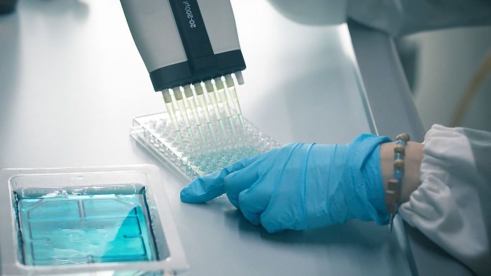
ELISA analysis is generally performed in 96 or 384-well polystyrene plates. These plates passively bind the proteins and antibodies, making ELISA analysis easy to design, develop and conduct. Besides, this specific binding facilitates the removal of unbound material during ELISA analysis. Moreover, the specificity acquired from washing the unbound materials makes immunoassay services focused on ELISA analysis for drug discovery and development studies. The current article is a step-by-step guide for ELISA analysis.
A guide for mastering ELISA analysis
Unless researchers have an alternative of an ELISA kit precoated with an antibody, the first step of ELISA analysis is the coating step. During this step, researchers coat the ELISA assay wells with the target antigen or antibody. The next step is to block all unbound sites using a blocking agent, followed by a series of washing steps and adding enzyme-conjugated antibodies. Post another washing step, a substrate is added that reacts with the enzyme to produce a colored signal.
As surface binding is the primary principle of ELISA assays, it requires several washing steps to remove any unbound material. This removal is critical to ensure that excess liquid is removed to prevent the dilution of solutions at every assay step. ELISA assays can become complex when assessing heterogeneous samples, for example, blood. However, irrespective of the sample type, detection is one of the most complex steps, requiring multiple antibodies to amplify the signal.
ELISA assays have numerous formats, including direct, indirect, sandwich, and competitive ELISA. The primary step of analyte immobilization is accomplished via capture antibodies or direct adsorption to the microplate. The detection can then be performed indirectly through enzyme-labeled secondary antibodies or directly through enzyme-labeled primary antibodies. Usually, the choice of enzyme substrates is horseradish peroxides or alkaline phosphatase. However, the final selection depends on the availability of instrumentation and required assay sensitivity.
Moreover, ELISA assays can generate three data outputs, quantitative, qualitative, and semi-quantitative. Quantitative ELISA incorporates data comparison with a standard curve to calculate analyte concentrations in study samples. On the other hand, qualitative ELISA involves achieving a yes or no answer indicating the presence of the analyte of interest in a study sample. Finally, semi-quantitative ELISA compares relative levels of the target analyte in study samples.
Usually, ELISA results are viewed by plotting a graph of optical density versus the log concentration. This graph produces a sigmoidal curve. Known concentrations of the analyte of interest are used to generate standard curves and measure unknown samples by comparing them with these standard curves. Besides, current ELISA plate readers have curve fitting software to determine the unknown concentrations directly from the graph. Importantly, robust ELISA primarily depends on a thorough ELISA assay validation.


